Home
/ Diagram Of Liver Fluke - Animal Biology Zoology Biology Parasitism 173 202 Life History Of The Sheep Liver Fluke The Egg Cell Is Produced In The Ovary Of The Fluke And Is Passed Into The Oviduct Where : Life cycle of liver fluke diagram.
Diagram Of Liver Fluke - Animal Biology Zoology Biology Parasitism 173 202 Life History Of The Sheep Liver Fluke The Egg Cell Is Produced In The Ovary Of The Fluke And Is Passed Into The Oviduct Where : Life cycle of liver fluke diagram.
Diagram Of Liver Fluke - Animal Biology Zoology Biology Parasitism 173 202 Life History Of The Sheep Liver Fluke The Egg Cell Is Produced In The Ovary Of The Fluke And Is Passed Into The Oviduct Where : Life cycle of liver fluke diagram.. In the uk the principle species is galba truncatula, the dwarf pond snail. Mode of transmission of liver fluke. Liver flukes and the environment. The diagram illustrates the four year treatment strategy demonstrated by parr and gray (2000) in which. Liver fluke disease is a chronic parasitic disease of the bile ducts.
They occur worldwide and range in size from about 5 millimetres (0.2 inch). Liver fluke is a collective name of a polyphyletic group of parasitic trematodes under the phylum platyhelminthes. In the continental u.s., fasciola hepatica blood chemistries suggestive of liver disease and eosinophilia support the diagnosis. Eblex suggests that liver fluke is often confused with poor nutrition, johne's disease, salmonellosis or parasitic gastroenteritis. If you live in an area where fluke prevalence is high, speak to your farm vet about forecasting and prevention of transmission.
A Well Labeled Pencil Sketch Diagram Of Liver Fluke Brainly In from hi-static.z-dn.net In the continental u.s., fasciola hepatica blood chemistries suggestive of liver disease and eosinophilia support the diagnosis. The most common types of liver flukes are clonorchis sinensis, opisthorchis viverrini and opisthorchis felineus. Liver flukes and the environment. Most of the damage is caused during the earliest stages of the parasite's development, as it travels through the animal's liver. Unlabeled digestive system diagram diagram human digestive system diagram unlabeled. Liver fluke life cycle liver fluke have an indirect life cycle involving a snail intermediate host. They are caused due to consumption of raw, undercooked, dried, or pickled freshwater fishes or by eating contaminated watercress. It is dorsoventrally flattened, oval in shape like a leaf and faint brownish in colour.
In the continental u.s., fasciola hepatica blood chemistries suggestive of liver disease and eosinophilia support the diagnosis.
Liver flukes infect the liver, gallbladder, and bile duct in humans. They occur worldwide and range in size from about 5 millimetres (0.2 inch). The diagram illustrates the four year treatment strategy demonstrated by parr and gray (2000) in which. Diagnosis of liver fluke is not simple. First diagram and second parts. Fasciola hepatica fasciolosis is an economically important and potentially fatal liver fluke in sheep. Recommendations for the control of liver flukes (fasciola hepatica) in cattle are based on strategically timed treatments with flukicidal. All three stages of liver fluke damage the liver and can cause clinical disease and production losses. Life cycle of liver fluke diagram. special collections, usda national agricultural library. Liver flukes are an important cause of acute and chronic disease in grazing sheep and cattle. They are principally parasites of the liver of various mammals, including humans. In the uk the principle species is galba truncatula, the dwarf pond snail. Liver flukes and the environment.
The southeast asian liver fluke (opisthorchis viverrini) chronically infects and affects tens of millions of people in regions of asia, leading to chronic illness and, importantly, inducing malignant cancer ( = cholangiocarcinoma). If you live in an area where fluke prevalence is high, speak to your farm vet about forecasting and prevention of transmission. First diagram and second parts. Undifferentiated fluke eggs are passed out in the faeces of infected animals and once washed out of the faeces. Liver fluke control involves treatment of infected animals, reduction of the.
Liver Fluke from image.slidesharecdn.com Liver flukes are an important cause of acute and chronic disease in grazing sheep and cattle. If producers are not normally affected they may not recognise the signs or treat routinely. Undifferentiated fluke eggs are passed out in the faeces of infected animals and once washed out of the faeces. It is dorsoventrally flattened, oval in shape like a leaf and faint brownish in colour. Recommendations for the control of liver flukes (fasciola hepatica) in cattle are based on strategically timed treatments with flukicidal. Caused by a flat worm called fasciola hepatica. Liver fluke disease is a chronic parasitic disease of the bile ducts. Unlabeled digestive system diagram diagram human digestive system diagram unlabeled.
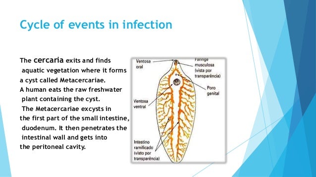
It is dorsoventrally flattened, oval in shape like a leaf and faint brownish in colour.
In spite of this, little is known, at the molecular level, about the parasite itself. Ingestion of fresh water plants with metacercaria or by drinking water with floating metacercariae. First diagram and second parts. In this article we will discuss about the external morphology of liver flukes. Liver flukes are an important cause of acute and chronic disease in grazing sheep and cattle. If you live in an area where fluke prevalence is high, speak to your farm vet about forecasting and prevention of transmission. The most common types of liver flukes are clonorchis sinensis, opisthorchis viverrini and opisthorchis felineus. Liver fluke, fasciola hepatica, is a highly pathogenic parasite which causes severe liver damage, especially in sheep, and can result in the sudden death of previously healthy animals. It is dorsoventrally flattened, oval in shape like a leaf and faint brownish in colour. Internal structure of liver fluke in blue with corresponding designations. While most infected persons do not show any symptoms, infections that last a long opisthorchis species are liver fluke parasites that humans can get by eating raw or undercooked fish, crabs, or crayfish from areas in asia and europe. Disease caused by liver fluke has increased in some european countries by up to 12 fold in recent years, and there is growing evidence to suggest it's increasing in the uk.1 the underlying cause appears to be climate change, favouring the survival and development of fluke stages that exist. There are more than 10,000 species of flukes.
Life cycle of liver fluke diagram. Eblex suggests that liver fluke is often confused with poor nutrition, johne's disease, salmonellosis or parasitic gastroenteritis. The life cycle of flukes is at first, liver flukes may cause no symptoms, or depending on the type and severity of the infection, they may cause fever, chills, abdominal pain, liver. Liver fluke disease is a chronic parasitic disease of the bile ducts. Unlabeled digestive system diagram diagram human digestive system diagram unlabeled.
Whole Mount In Situ Hybridization Of Fhtlm In Adult Liver Fluke A Download Scientific Diagram from www.researchgate.net Find stockbilleder af fasciola hepatica internal structure liver fluke i hd og millionvis af andre royaltyfri stockbilleder, illustrationer og vektorer i shutterstocks samling. First diagram and second parts. Liver fluke control involves treatment of infected animals, reduction of the. Mode of transmission of liver fluke. They occur worldwide and range in size from about 5 millimetres (0.2 inch). They are caused due to consumption of raw, undercooked, dried, or pickled freshwater fishes or by eating contaminated watercress. The most common types of liver flukes are clonorchis sinensis, opisthorchis viverrini and opisthorchis felineus. If you live in an area where fluke prevalence is high, speak to your farm vet about forecasting and prevention of transmission.
Diagnosis of liver fluke is not simple.
Fasciola hepatica (the common liver fluke or sheep liver fluke), which causes fascioliasis and typically infects sheep and cattle. First diagram and second parts. Unlabeled digestive system diagram diagram human digestive system diagram unlabeled. Find stockbilleder af fasciola hepatica internal structure liver fluke i hd og millionvis af andre royaltyfri stockbilleder, illustrationer og vektorer i shutterstocks samling. For a long time, the agent of opisthorchiasis, a widespread parasitic disease caused by eating infected fish, was mainly the object of medical and parasitological studies. The southeast asian liver fluke (opisthorchis viverrini) chronically infects and affects tens of millions of people in regions of asia, leading to chronic illness and, importantly, inducing malignant cancer ( = cholangiocarcinoma). If producers are not normally affected they may not recognise the signs or treat routinely. In the uk the principle species is galba truncatula, the dwarf pond snail. Undifferentiated fluke eggs are passed out in the faeces of infected animals and once washed out of the faeces. Learn vocabulary, terms and more with flashcards, games and other study tools. Diagnosis of liver fluke is not simple. Disease caused by liver fluke has increased in some european countries by up to 12 fold in recent years, and there is growing evidence to suggest it's increasing in the uk.1 the underlying cause appears to be climate change, favouring the survival and development of fluke stages that exist. They occur worldwide and range in size from about 5 millimetres (0.2 inch).
The diagram illustrates the four year treatment strategy demonstrated by parr and gray (2000) in which diagram of liver. Learn vocabulary, terms and more with flashcards, games and other study tools.

